Encapsulating peritoneal sclerosis is a rare but serious condition that can occur in patients, most commonly those undergoing long-term peritoneal dialysis. It is characterized by progressive fibrosis and thickening of the peritoneum, which leads to encapsulation of the small intestine and symptoms of bowel obstruction. Radiology plays an essential role in identifying this condition at different stages and guiding clinical management. Understanding how encapsulating peritoneal sclerosis appears on imaging studies helps clinicians make an early diagnosis, monitor disease progression, and plan appropriate treatment options. Because of its nonspecific clinical presentation, radiology often provides the key insights needed for accurate detection.
Overview of Encapsulating Peritoneal Sclerosis
Encapsulating peritoneal sclerosis (EPS) is defined by the formation of a dense fibrotic membrane that surrounds the bowel loops, often leading to impaired motility and obstruction. The disease can progress slowly and may remain undetected until patients develop severe abdominal pain, nausea, vomiting, or recurrent intestinal blockages. Risk factors include long-term peritoneal dialysis, recurrent peritonitis, and the use of high-glucose dialysate solutions. Although EPS is uncommon, it is recognized as one of the most severe complications of peritoneal dialysis, making radiology crucial in its detection and evaluation.
Importance of Radiology in EPS
Radiology is the cornerstone of diagnosing encapsulating peritoneal sclerosis. While clinical history and physical examination provide initial clues, imaging helps reveal structural changes in the peritoneum and bowel. Early findings may be subtle, so radiologists must be familiar with the specific imaging features of EPS. Different radiological modalities, including computed tomography (CT), magnetic resonance imaging (MRI), and ultrasound, can be used to evaluate the severity and progression of the condition. CT remains the most commonly used imaging technique because it provides detailed visualization of the abdomen and pelvis.
Radiological Features on CT
Computed tomography is considered the gold standard for diagnosing EPS. Several key findings are often described
- Bowel wall thickeningSmall bowel loops may show diffuse or localized thickening, suggesting fibrosis and inflammation.
- Peritoneal thickening and calcificationThe peritoneum may appear irregular, with focal or diffuse calcifications that point toward chronic inflammation.
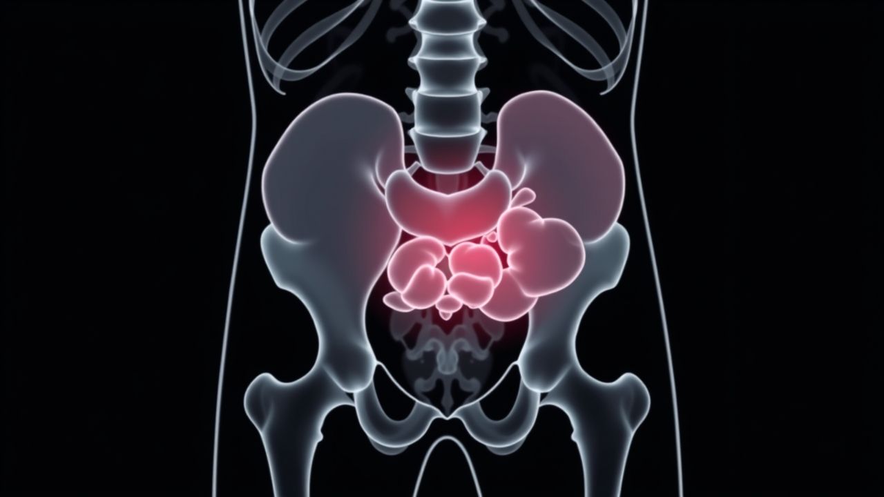
- Encapsulation of bowel loopsThe small intestine may appear clumped together, encased by a fibrous membrane resembling a cocoon.
- Loculated fluid collectionsAscites or localized fluid pockets may be present, complicating the clinical picture.
- Signs of obstructionDilated bowel loops proximal to the fibrotic segments may indicate mechanical obstruction caused by encapsulation.
Role of MRI in Encapsulating Peritoneal Sclerosis
Magnetic resonance imaging provides excellent soft tissue contrast and can be used to assess the peritoneum and bowel without radiation exposure. MRI may reveal similar findings to CT, including peritoneal thickening, bowel encapsulation, and fibrosis. In addition, MRI can evaluate peritoneal enhancement patterns after contrast administration, which may reflect ongoing inflammation. Although MRI is not always the first-line imaging choice due to cost and accessibility, it serves as a valuable complementary tool in complex cases or in patients who cannot undergo CT scans.
Ultrasound Findings in EPS
Ultrasound is a readily available imaging modality that may show abnormal thickening of the bowel wall, dilated fluid-filled loops, and echogenic strands representing fibrotic tissue. However, ultrasound is operator-dependent and less sensitive than CT in detecting encapsulating membranes. It is often used as an initial imaging study but usually requires confirmation with cross-sectional imaging.
Radiology in Differential Diagnosis
EPS can mimic other abdominal conditions such as peritoneal carcinomatosis, tuberculous peritonitis, or sclerosing mesenteritis. Radiology helps differentiate EPS from these conditions by highlighting distinctive features such as calcified peritoneal thickening and encapsulated bowel loops. Correlation with clinical history, especially prior peritoneal dialysis, is essential for an accurate diagnosis. Identifying these radiological patterns prevents misdiagnosis and allows timely initiation of appropriate therapy.
Staging and Severity Assessment
Radiology not only aids in diagnosis but also helps stage the severity of encapsulating peritoneal sclerosis. CT scans can classify the disease into early, intermediate, and late stages
- Early stageSubtle peritoneal thickening with minimal bowel changes.
- Intermediate stageProgressive fibrosis, encapsulation, and moderate bowel obstruction.
- Late stageAdvanced encapsulation with dense fibrotic membranes, significant obstruction, and extensive calcification.
Understanding the stage of the disease helps clinicians decide between conservative management, immunosuppressive therapy, or surgical intervention.
Clinical Correlation with Radiology
Radiological findings must always be interpreted in conjunction with patient symptoms and medical history. For example, a patient undergoing long-term peritoneal dialysis who develops recurrent abdominal pain and weight loss should be evaluated for EPS. Radiology confirms the suspicion, and early diagnosis may prevent severe complications. Clinical-radiological correlation is critical because some findings, such as peritoneal thickening, may overlap with other conditions.
Limitations of Radiological Imaging
Despite its importance, radiology has limitations in EPS. Some early changes may be too subtle to detect, leading to delayed diagnosis. Calcifications and thickening seen on CT may not always correlate with disease severity. Moreover, advanced disease may present challenges in distinguishing EPS from malignant or infectious conditions. Radiologists must therefore approach interpretation with caution and integrate findings with the patient’s clinical background.
Impact of Radiology on Treatment Planning
Radiological assessment directly influences treatment decisions in encapsulating peritoneal sclerosis. Early detection allows for timely cessation of peritoneal dialysis and initiation of medical therapy such as corticosteroids or tamoxifen. In advanced cases, surgery may be necessary to remove the fibrotic capsule and release the bowel loops. Radiology guides surgeons by identifying the extent of encapsulation and highlighting potential complications before intervention. Accurate imaging thus plays a central role in optimizing outcomes for patients with EPS.
Future Perspectives in Radiology for EPS
Advances in imaging technology continue to improve the evaluation of encapsulating peritoneal sclerosis. High-resolution CT, diffusion-weighted MRI, and functional imaging techniques may provide new insights into disease activity and response to therapy. Research is ongoing to refine radiological criteria for early detection and to differentiate EPS from similar peritoneal conditions more reliably. These advancements hold promise for earlier diagnosis and better patient outcomes.
Encapsulating peritoneal sclerosis is a rare but severe complication of long-term peritoneal dialysis, and radiology remains the most valuable tool in its diagnosis and management. CT, MRI, and ultrasound each provide unique contributions to identifying characteristic features such as peritoneal thickening, calcification, and bowel encapsulation. By staging disease progression and distinguishing EPS from other conditions, radiology guides clinical decisions and improves treatment planning. Ongoing advances in imaging technology promise to enhance early detection and support better care for patients affected by this challenging disorder.