Pleural effusion is a medical condition characterized by the accumulation of excess fluid in the pleural space, the thin cavity between the lungs and the chest wall. Radiological imaging plays a critical role in detecting, diagnosing, and managing pleural effusions. Accurate interpretation of these images allows clinicians to determine the type, size, and underlying cause of the effusion, guiding appropriate treatment strategies. Understanding the radiological appearance of pleural effusion, the types of imaging modalities used, and the clinical significance of various findings is essential for both healthcare professionals and patients seeking insight into this condition.
Understanding Pleural Effusion
Pleural effusion can result from a wide range of medical conditions, including heart failure, pneumonia, malignancies, pulmonary embolism, and liver or kidney disease. The fluid accumulation can impair lung expansion, leading to shortness of breath, chest pain, and cough. Radiological imaging not only confirms the presence of pleural effusion but also helps distinguish between transudative and exudative types, which is essential for determining the underlying cause.
Common Symptoms Indicating the Need for Imaging
- Shortness of breath or difficulty breathing
- Sharp or dull chest pain, particularly with deep breaths
- Persistent cough
- Decreased breath sounds on physical examination
- Signs of systemic disease, such as fever or weight loss
Types of Radiological Imaging for Pleural Effusion
Various imaging modalities are used to evaluate pleural effusions, each providing unique insights into the presence, volume, and characteristics of the fluid.
Chest X-Ray
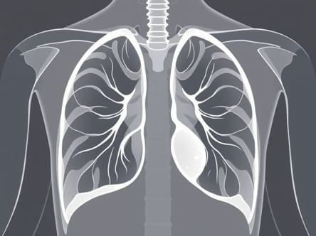
Chest X-ray is the most common initial imaging modality for detecting pleural effusions. It is widely available, quick, and cost-effective. On a chest X-ray, pleural effusion typically appears as a homogeneous opacity at the lung bases with blunting of the costophrenic angles. Larger effusions may cause a meniscus sign, a curved upper margin indicating fluid accumulation. Supine or lateral decubitus X-rays may be employed for patients unable to stand, which helps detect smaller or loculated effusions.
Ultrasound
Ultrasound imaging is highly sensitive for detecting pleural effusions and is particularly useful for guiding thoracentesis, a procedure to drain fluid from the pleural space. Ultrasound can distinguish between free-flowing and loculated effusions and may help identify septations, fibrin strands, or pleural thickening. This modality is radiation-free, portable, and provides real-time guidance for safe and effective fluid removal.
Computed Tomography (CT) Scan
CT scans provide detailed cross-sectional images of the chest, offering superior visualization of pleural effusions and associated structures. CT imaging can detect even small amounts of fluid, assess underlying lung pathology, and identify complications such as empyema or pleural tumors. It is particularly useful in complex cases where chest X-ray findings are inconclusive or when malignancy or infection is suspected.
Radiological Features of Pleural Effusion
Radiological images provide distinct features that help identify pleural effusions and guide diagnosis. Recognizing these features is essential for accurate interpretation and appropriate clinical management.
Chest X-Ray Findings
- Blunted Costophrenic Angles One of the earliest signs of pleural effusion.
- Meniscus Sign Curved upper border of the fluid, often seen in larger effusions.
- Homogeneous Opacity Fluid appears as a dense area on the X-ray.
- Contralateral Mediastinal Shift In massive effusions, the mediastinum may shift away from the affected side.
- Layering in Supine Position Effusions may layer posteriorly, visible on lateral decubitus films.
Ultrasound Findings
- Anechoic or Hypoechoic Fluid Represents free-flowing pleural fluid.
- Septations Indicate loculated effusions, often associated with infection or hemothorax.
- Dynamic Changes Real-time imaging shows fluid movement during respiration.
- Guided Needle Placement Ultrasound can direct thoracentesis safely.
CT Scan Findings
- High-Resolution Detection Small or complex effusions are easily identified.
- Pleural Thickening or Nodularity May suggest malignancy or chronic inflammation.
- Associated Lung Changes Consolidation, atelectasis, or masses can be evaluated concurrently.
- Empyema Identification CT can detect infected or loculated pleural fluid collections.
Clinical Significance of Radiological Imaging
Radiological imaging plays a critical role in determining the etiology, extent, and severity of pleural effusion. By analyzing the size, density, and distribution of fluid, healthcare providers can differentiate between transudative and exudative effusions, which informs further diagnostic testing and treatment strategies. Imaging also assists in monitoring response to therapy, such as fluid reduction after diuretics in heart failure or resolution of infection following antibiotics.
Guiding Therapeutic Interventions
Imaging modalities, particularly ultrasound and CT scans, are invaluable for guiding therapeutic procedures like thoracentesis or chest tube placement. Proper visualization ensures accurate needle insertion, minimizes complications, and allows for complete fluid drainage. This targeted approach improves patient outcomes and reduces procedure-related risks.
Limitations and Considerations
While radiological imaging is essential in managing pleural effusion, there are limitations to consider. Chest X-rays may miss small or loculated effusions, especially in supine patients. Ultrasound is operator-dependent, and CT scans involve radiation exposure and may require contrast agents. Clinical correlation with patient history, physical examination, and laboratory findings remains essential for accurate diagnosis and treatment planning.
Integration with Clinical Assessment
- Correlate imaging findings with patient symptoms and physical examination.
- Use imaging to monitor progression or resolution of effusion over time.
- Combine radiological findings with laboratory analysis of pleural fluid when necessary.
- Consider patient-specific factors such as mobility, comorbidities, and procedural risks.
Radiological imaging is a cornerstone in the diagnosis and management of pleural effusions. From chest X-rays to ultrasound and CT scans, each modality provides unique information that helps clinicians identify the presence, type, and underlying cause of fluid accumulation. Understanding radiological features such as blunted costophrenic angles, meniscus signs, septations, and pleural thickening allows for accurate interpretation and informed decision-making. Imaging also plays a vital role in guiding therapeutic interventions, monitoring treatment response, and ensuring patient safety. By integrating radiological findings with clinical assessment, healthcare providers can deliver comprehensive, effective care for individuals affected by pleural effusions.
Ultimately, the radiological image of pleural effusion serves as an essential diagnostic tool, providing clarity and direction in the management of a condition that can impact respiratory function and overall health. Accurate interpretation, combined with clinical expertise, ensures timely and appropriate interventions, improving patient outcomes and enhancing quality of life for those affected by pleural fluid accumulation.