The maxillary second premolar is one of the most discussed teeth in dental anatomy because of its variability and importance in chewing, stability, and overall oral function. Many people become curious about how many roots this tooth has, especially when learning about dental treatments such as root canals or extractions. Although it may seem like a simple question, the anatomy of this tooth can vary from person to person. Understanding these variations helps patients and professionals appreciate why careful examination is crucial before performing any dental procedure. Exploring its structure provides deeper insight into dental health and tooth morphology.
General Anatomy of the Maxillary Second Premolar
The maxillary second premolar is located behind the first premolar and in front of the first molar, positioned in the upper jaw. It plays an important role in grinding food and maintaining facial structure. Its shape and root configuration differ slightly from the first premolar, but both contribute significantly to chewing efficiency.
While the crown anatomy is relatively consistent, root morphology shows greater variation. Many dental professionals rely on imaging techniques to assess the number of roots and root canals before performing treatment, as misinterpreting the structure can lead to complications.
How Many Roots Does the Maxillary Second Premolar Usually Have?
In most people, the maxillary second premolar typically has one root. This is the most common anatomical configuration and is considered the standard pattern in dental studies. Despite this general rule, variations do occur, making this tooth one of the more complex premolars to evaluate.
The most frequently reported root pattern includes
- One root (most common)
- Two roots (less common)
- Rare multi-rooted variations
Though one root is dominant, the number of root canals within that root can also vary, adding another layer of complexity to its anatomy.
One-Root Morphology
Most maxillary second premolars contain a single root that is slender, slightly flattened, and often conical. This single root may house one or two root canals. Dentists often expect one canal but prepare for the possibility of two because of the internal complexity.
One Canal
When the tooth contains one canal, the canal usually runs straight to the apex. This configuration simplifies endodontic treatments because there is usually only one pathway to clean and fill.
Two Canals in One Root
It is also possible for a single root to contain two canals. These canals may merge before reaching the apex or remain separate throughout their length. This arrangement is more challenging to treat due to increased curvature and the potential for branching.
Two-Root Variations
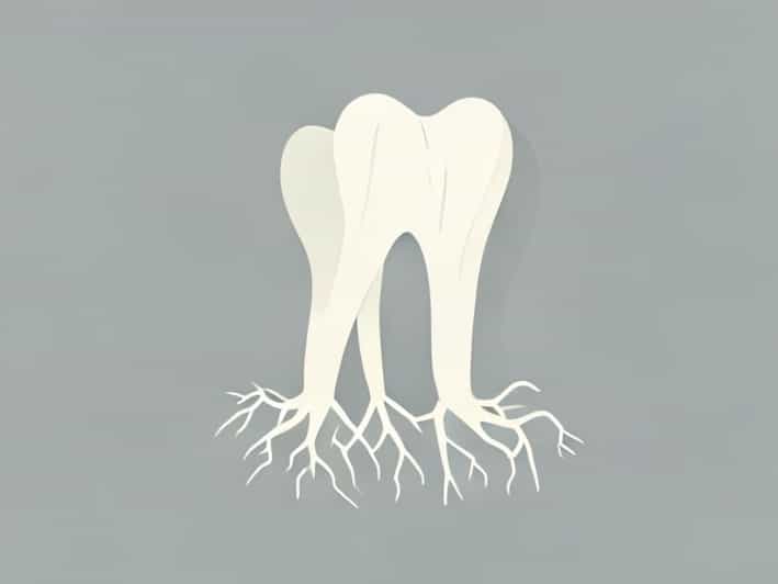
The maxillary second premolar may also present with two separate roots, though this is much less common. When two roots exist, they are usually positioned as a buccal (cheek-side) and palatal (tongue-side) root, similar to the configuration of the first maxillary premolar.
Characteristics of Two-Rooted Premolars
Two-rooted variants may include
- A buccal root with one canal
- A palatal root with one canal
- Occasional branching within either root
This configuration is important to identify before treatment because failing to detect the second root can leave a canal untreated, increasing the risk of later infection.
Rare Multi-Rooted Cases
Though extremely rare, the maxillary second premolar can sometimes have three roots. This is a rare anomaly and often resembles the structure of a small molar. The roots may include
- Two buccal roots
- One palatal root
These teeth almost always contain multiple canals, making endodontic treatment significantly more complex. Advanced imaging, such as cone beam CT scanning, is often recommended for identifying such variations.
Factors Influencing Root Variations
Several factors contribute to root variation in the maxillary second premolar. Although genetics play a major role, environmental influences and developmental factors may also affect how the roots form.
Genetic Influence
Root morphology can vary among populations based on hereditary traits. Certain groups may show a higher likelihood of having two roots or multiple canals.
Developmental Factors
The shape and number of roots develop early during tooth formation. If the root sheath splits or folds differently, the number of roots can change.
Individual Variation
No two people are exactly alike, and natural variation means that even within the same family, root structures can differ. Dentists always consider this when diagnosing a tooth.
Clinical Importance of Knowing Root Anatomy
Understanding how many roots a maxillary second premolar has is essential for safe and effective dental treatment. Root anatomy affects several procedures, including root canals, extractions, and orthodontic planning.
Endodontic Treatment
Root canal procedures require precise cleaning of all canals. Missing a canal is one of the most common causes of treatment failure. Because this premolar can sometimes contain multiple canals, careful imaging and exploration are crucial.
Dental Extractions
Extractions are easier when root anatomy is understood beforehand. A single root usually allows for smoother extraction, while multiple roots may increase resistance and require different surgical techniques.
Orthodontic Considerations
Root length, curvature, and number influence tooth movement. Knowing whether a tooth has multiple roots helps orthodontists avoid undue force that could damage the root structure.
Diagnostic Methods for Identifying Root Number
Dentists use various tools to identify the number of roots in a maxillary second premolar. These diagnostic methods help ensure accuracy before any major treatment.
Traditional X-Rays
Standard dental radiographs provide useful information about root shape and number. However, overlapping structures may sometimes make interpretation difficult.
Cone Beam Computed Tomography (CBCT)
CBCT scans produce three-dimensional images, offering a clear view of the roots and canals. This method is the most reliable for analyzing complex root morphology.
Clinical Examination
During root canal treatment, the dentist may discover additional canals by exploring the pulp chamber. Experience and technique play an important role in locating hidden canals.
Why Variation Matters
Root variation in the maxillary second premolar is more than an interesting anatomical fact-it has real implications for dental health and treatment success. A tooth with more roots or canals is not weaker or defective; it simply requires thoughtful evaluation. Understanding these variations ensures proper diagnosis, reduces risk, and helps maintain long-term oral health.
The maxillary second premolar typically has one root, making it simpler than many other upper teeth. However, variations do exist, including the possibility of two roots or, in rare cases, three. The number of canals within each root can also differ, adding complexity to dental treatments. By recognizing these anatomical differences, dental professionals can plan more effective procedures, and patients can better understand the importance of detailed dental assessments. Root anatomy in this tooth highlights the fascinating diversity of the human body and its intricate design.