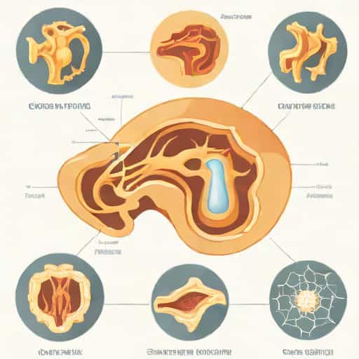
Endochondral ossification is a fundamental biological process through which most of the bones in the human body develop. It is particularly responsible for forming long bones such as the femur, tibia, and humerus. This process begins with a cartilage template that is gradually replaced by bone tissue. Understanding the steps of endochondral ossification is essential for students of anatomy, physiology, and medicine, as it provides insight into bone growth, skeletal development, and repair mechanisms. Each step in the process is highly regulated and involves a series of cellular and molecular changes that ensure proper bone formation.
Definition of Endochondral Ossification
Endochondral ossification is a type of bone formation in which the bone develops by replacing a pre-existing hyaline cartilage model. This method of ossification is distinct from intramembranous ossification, which forms bones directly from mesenchymal tissue. Endochondral ossification plays a crucial role during fetal development and continues postnatally to allow for longitudinal growth of long bones until adulthood. It also contributes to the healing of bone fractures through cartilage formation followed by ossification.
Importance of Endochondral Ossification
- Facilitates the growth of long bones during childhood and adolescence.
- Supports proper skeletal structure and shape.
- Enables repair of bone injuries through cartilage intermediates.
- Ensures the development of joints and epiphyseal plates for movement.
Step 1 Formation of the Cartilage Model
The process of endochondral ossification begins with the formation of a cartilage model made of hyaline cartilage. Mesenchymal cells aggregate in the shape of the future bone and differentiate into chondrocytes. These chondrocytes produce a cartilaginous matrix that outlines the general structure of the future bone. The cartilage model is avascular, meaning it lacks blood vessels, and is surrounded by a perichondrium, which provides some structural support and nourishment.
Key Points of Cartilage Model Formation
- Mesenchymal cells differentiate into chondrocytes.
- Chondrocytes secrete the cartilage matrix.
- The model establishes the shape and size of the future bone.
- Perichondrium surrounds the cartilage to protect and support growth.
Step 2 Growth of the Cartilage Model
Once the cartilage model is formed, it grows in size through two mechanisms interstitial growth and appositional growth. Interstitial growth occurs as chondrocytes divide and secrete matrix from within, expanding the cartilage internally. Appositional growth occurs when new chondrocytes and matrix are added to the surface of the cartilage model. During this phase, the cartilage continues to elongate, setting the stage for ossification in the center.
Features of Cartilage Growth
- Chondrocytes undergo division and produce more matrix.
- Interstitial growth increases the internal volume of the cartilage.
- Appositional growth adds layers of cartilage to the periphery.
- Prepares the model for the primary ossification center.
Step 3 Formation of the Primary Ossification Center
The primary ossification center forms in the central region of the cartilage model, usually in the diaphysis or shaft of the future bone. In this step, the perichondrium surrounding the cartilage differentiates into a periosteum, and osteoblasts begin to replace cartilage with bone. Blood vessels invade the cartilage, bringing nutrients and osteogenic cells that facilitate the transformation of cartilage into bone. Chondrocytes in the center hypertrophy and secrete enzymes that calcify the surrounding matrix, which eventually leads to cell death and formation of cavities for new bone tissue.
Important Events in Primary Ossification
- Perichondrium transforms into periosteum.
- Osteoblasts invade and deposit bone matrix.
- Chondrocytes hypertrophy and calcify the surrounding cartilage.
- Blood vessels penetrate to supply osteogenic cells.
- Cavities form as chondrocytes die, preparing for bone deposition.
Step 4 Development of the Medullary (Marrow) Cavity
As ossification progresses, the diaphysis becomes hollowed out to form the medullary cavity. Osteoclasts resorb the calcified cartilage and bone in the center, creating space for bone marrow. This cavity will eventually house red and yellow marrow, which are essential for hematopoiesis and fat storage, respectively. This step ensures that long bones are both strong and lightweight, allowing them to support the body without excessive weight.
Features of Medullary Cavity Formation
- Osteoclasts resorb the central calcified cartilage.
- Bone marrow occupies the newly formed cavity.
- Diaphysis becomes a tubular structure with cortical bone walls.
- Supports both structural strength and functional hematopoiesis.
Step 5 Formation of Secondary Ossification Centers
Secondary ossification centers appear in the epiphyses, or ends, of the long bones, usually after birth. These centers develop similarly to the primary center but do not form a medullary cavity. Instead, spongy bone is deposited, while cartilage remains at the epiphyseal plates and joint surfaces. The epiphyseal cartilage is crucial for longitudinal growth during childhood and adolescence. Over time, the secondary ossification centers ossify, leaving only the articular cartilage and the epiphyseal plate.
Significance of Secondary Ossification
- Forms the ends of long bones and joint surfaces.
- Maintains cartilage for continued growth at epiphyseal plates.
- Contributes to bone lengthening until adulthood.
- Ensures proper formation of spongy bone at epiphyses.
Step 6 Formation of Epiphyseal Plates
The epiphyseal plates, also called growth plates, remain as regions of cartilage between the primary and secondary ossification centers. These plates allow the bones to grow in length while maintaining structural integrity. Chondrocytes at the growth plates continue to divide, expand, and eventually ossify, contributing to the longitudinal growth of bones. Once the individual reaches adulthood, the epiphyseal plates ossify completely, resulting in the cessation of bone growth.
Functions of Epiphyseal Plates
- Facilitate longitudinal growth of long bones.
- Provide flexibility for bone development during childhood and adolescence.
- Ensure proper alignment and shape of the bone ends.
- Eventually ossify to form the epiphyseal line in adults.
Step 7 Completion of Endochondral Ossification
Endochondral ossification is considered complete once the cartilage is replaced by bone, the epiphyseal plates ossify, and the bone achieves its mature length and shape. At this stage, bones are fully capable of providing support, leverage, and protection. However, the process of bone remodeling continues throughout life, adjusting bone structure in response to mechanical stress and repair needs.
Summary of the Steps
- Formation of cartilage model
- Growth of cartilage model (interstitial and appositional)
- Formation of primary ossification center in the diaphysis
- Development of medullary cavity by osteoclast activity
- Formation of secondary ossification centers in epiphyses
- Maintenance of epiphyseal plates for longitudinal growth
- Completion of ossification and maturation of the bone
The steps of endochondral ossification outline the complex yet highly organized process of bone development in long bones. From the formation of the cartilage template to the establishment of primary and secondary ossification centers, this process ensures proper bone growth, structure, and function. Understanding each step is crucial for students of anatomy, medicine, and biology, as it provides insight into skeletal development, growth disorders, and the healing of fractures. Endochondral ossification exemplifies the intricate coordination between cellular activity, cartilage formation, and bone deposition, highlighting the marvel of human skeletal development.