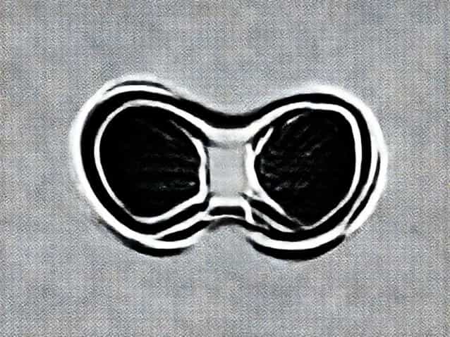
Biconcave deformity of the vertebral bodies is a notable spinal abnormality that can affect individuals of various ages and often signifies underlying health issues. This condition is characterized by a concave depression on both the superior and inferior surfaces of the vertebral bodies, giving them a distinct fish-mouth appearance on radiological imaging. Biconcave vertebral deformities may develop gradually and are frequently associated with metabolic bone disorders, chronic illnesses, or congenital conditions. Recognizing this deformity is critical for early diagnosis, appropriate management, and prevention of further complications such as spinal instability, pain, or neurological symptoms.
Understanding the Anatomy and Pathophysiology
The vertebral bodies are the thick, cylindrical portions of the vertebrae that provide structural support to the spinal column and bear the majority of axial load. In a healthy spine, these bodies maintain a relatively uniform, rectangular shape. Biconcave deformity occurs when both the upper (superior) and lower (inferior) endplates of a vertebral body collapse or weaken, leading to concave indentations. This abnormal shape can compromise the structural integrity of the spine and alter normal biomechanics, which may contribute to chronic back pain or increased susceptibility to further fractures.
Causes of Biconcave Vertebral Deformity
The development of biconcave vertebral deformities is typically linked to conditions that weaken the bone or disrupt normal vertebral growth. Some common causes include
- OsteoporosisReduced bone mineral density weakens vertebral bodies, making them susceptible to compression and biconcave changes, especially in older adults.
- OsteomalaciaDeficient vitamin D or phosphate metabolism can cause softening of the vertebral bones, leading to endplate depression.
- ThalassemiaChronic anemia and bone marrow expansion in thalassemia patients can lead to characteristic biconcave vertebral deformities, often referred to as fish vertebrae.
- Multiple MyelomaMalignant infiltration of the bone marrow may weaken vertebral structures, resulting in endplate concavity.
- Congenital or developmental disordersCertain skeletal dysplasias or genetic disorders can predispose individuals to abnormal vertebral shapes, including biconcave deformities.
- TraumaRepeated or severe mechanical stress can contribute to compression of vertebral endplates in susceptible individuals.
Clinical Presentation
The presentation of biconcave vertebral deformities can vary depending on the underlying cause and severity of the condition. Some individuals remain asymptomatic and the deformity is discovered incidentally during imaging for other reasons. Symptomatic cases may present with
- Chronic back pain or discomfort localized to the affected vertebral segments.
- Loss of height due to multiple vertebral body collapses.
- Postural changes such as kyphosis or spinal curvature abnormalities.
- Neurological symptoms if the spinal cord or nerve roots are compressed, including numbness, tingling, or weakness in the limbs.
- Fatigue or malaise in systemic conditions like anemia or chronic metabolic disorders.
Diagnostic Approaches
Accurate diagnosis of biconcave vertebral deformities requires imaging and, in some cases, laboratory investigations to determine the underlying cause. Common diagnostic methods include
- X-rayRadiographs can clearly reveal the biconcave fish-mouth appearance of affected vertebrae and help assess the extent of deformity.
- CT ScanComputed tomography provides detailed images of bone structures and can help differentiate biconcave deformities from fractures or other spinal anomalies.
- MRIMagnetic resonance imaging evaluates both bone and soft tissue, including intervertebral discs and spinal cord, which is useful if neurological symptoms are present.
- Bone density testsDual-energy X-ray absorptiometry (DEXA) may be used to assess osteoporosis or other bone mineral deficiencies.
- Laboratory investigationsBlood tests for calcium, phosphate, vitamin D, and markers of bone turnover can help identify metabolic or systemic causes.
Management and Treatment
Management of biconcave vertebral deformities depends on the underlying etiology, severity of symptoms, and risk of progression. General approaches include
Medical Management
- Osteoporosis Bisphosphonates, vitamin D, calcium supplementation, and lifestyle modifications like weight-bearing exercises.
- Osteomalacia Correction of vitamin D and phosphate deficiencies through dietary supplementation or medications.
- Thalassemia Regular transfusions, iron chelation therapy, and management of bone health through supplements and monitoring.
- Multiple Myeloma Chemotherapy, targeted therapies, and supportive bone-strengthening treatments.
Physical Therapy and Lifestyle Adjustments
Physical therapy can help maintain spinal mobility, strengthen core muscles, and reduce the risk of further vertebral collapse. Postural training, safe lifting techniques, and ergonomic adjustments may also alleviate symptoms and improve quality of life.
Surgical Interventions
In severe cases with progressive deformity, instability, or neurological compromise, surgical options may be considered. Procedures can include vertebral augmentation (vertebroplasty or kyphoplasty) to restore height and stability, or spinal fusion for structural support. Surgery is generally reserved for cases unresponsive to conservative therapy or where neurological deficits are present.
Prognosis
The prognosis for individuals with biconcave vertebral deformities varies according to cause and treatment efficacy. With early detection and proper management, progression can be slowed, pain can be reduced, and quality of life can be maintained. In cases related to chronic systemic diseases, ongoing monitoring and comprehensive care are essential to prevent further complications. Untreated or severe deformities may result in chronic pain, spinal instability, or neurological deficits.
Preventive Measures
Prevention of biconcave vertebral deformities primarily focuses on maintaining bone health and addressing risk factors
- Ensure adequate intake of calcium and vitamin D.
- Engage in regular weight-bearing and resistance exercises.
- Avoid smoking and excessive alcohol consumption.
- Monitor and manage chronic illnesses that affect bone health, such as diabetes, kidney disease, or blood disorders.
- Regular screening for osteoporosis or metabolic bone disorders in at-risk populations.
Early Recognition
Early recognition and intervention are crucial. Routine imaging for individuals with known risk factors or unexplained back pain can help detect vertebral deformities before complications arise. Prompt diagnosis allows for timely treatment of the underlying cause, minimizing the risk of progression.
Biconcave deformity of the vertebral bodies is a significant clinical finding that reflects underlying bone weakness or systemic disease. Understanding its causes, clinical presentation, and diagnostic methods is essential for appropriate management. Treatment strategies range from medical therapy and lifestyle adjustments to surgical intervention in severe cases. Early detection, preventive measures, and comprehensive care can improve patient outcomes, reduce pain, and prevent further spinal complications. By maintaining awareness of risk factors and prioritizing bone health, individuals can minimize the impact of this deformity and maintain spinal function over time.