Undergoing an X-ray examination can be a routine part of medical care, but many patients are often unsure about what to expect from the procedure. An X-ray examination procedure is a safe, non-invasive imaging technique used to visualize the internal structures of the body. It relies on a controlled dose of radiation to create images of bones, organs, and other tissues, helping healthcare providers diagnose conditions ranging from fractures and infections to lung diseases and dental issues. Understanding the steps involved, preparation, and safety measures can help patients feel more comfortable and informed during the process.
What is an X-Ray Examination?
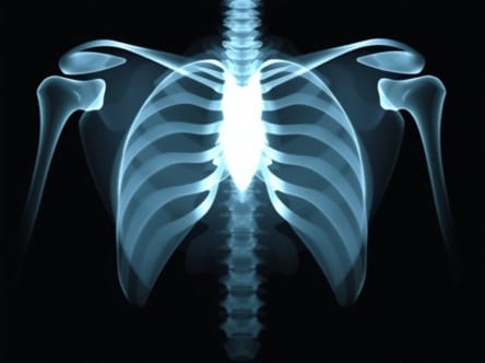
An X-ray examination, also known as radiography, is a diagnostic tool that uses electromagnetic radiation to produce images of the internal structures of the body. X-rays pass through soft tissues and are absorbed by denser materials like bones, producing clear contrast in the resulting images. These images are then analyzed by radiologists or other healthcare professionals to identify abnormalities or injuries. X-rays are widely used in medicine due to their speed, efficiency, and effectiveness in providing critical diagnostic information.
Preparation Before the X-Ray
Preparing for an X-ray examination is generally straightforward, though certain procedures may require specific instructions. Common preparation steps include
- Removing metal objects such as jewelry, watches, and belts that can interfere with image clarity.
- Wearing a hospital gown or clothing that does not contain metallic components.
- Informing the technician if you are pregnant or suspect pregnancy, as precautions may be necessary to protect the fetus.
- Following any dietary restrictions or instructions if the X-ray involves contrast materials or specific body areas like the gastrointestinal tract.
By following these preparation steps, patients can ensure the X-ray images are as accurate as possible and minimize the need for repeat scans.
The X-Ray Examination Procedure
The procedure itself is generally quick and painless. The exact steps may vary depending on the body part being examined, but the core process includes several key elements
Positioning
The technician will guide the patient into the appropriate position to obtain the clearest images. This may involve standing, sitting, or lying on an X-ray table. Proper positioning is critical for accurate imaging and may require the use of supports or immobilization devices to keep the patient still during the scan.
Use of Protective Shields
To minimize unnecessary exposure to radiation, protective shields made of lead may be placed over parts of the body that are not being imaged. These shields are particularly important for reproductive organs and other sensitive areas. Patients should remain still while the shield is in place to ensure safety and image quality.
Image Capture
The X-ray machine emits a controlled beam of radiation directed toward the targeted area. Depending on the body part, multiple images may be taken from different angles. The patient may be asked to hold their breath briefly to reduce motion blur, especially for chest or abdominal X-rays. The exposure itself only lasts a few seconds, though the overall procedure may take 10 to 30 minutes depending on complexity.
Types of X-Ray Examinations
X-ray examinations can vary widely based on the body part and medical purpose. Some common types include
- Chest X-RayUsed to evaluate lungs, heart, and chest wall for conditions like pneumonia, heart failure, or fractures.
- Bone X-RayHelps detect fractures, dislocations, infections, and bone density issues.
- Dental X-RayProvides images of teeth, jawbones, and oral structures to diagnose cavities, impacted teeth, or gum disease.
- Abdominal X-RayEvaluates organs such as the stomach, intestines, liver, and kidneys for blockages, stones, or other abnormalities.
- Contrast X-RayUses a special dye or contrast material to enhance visibility of specific organs or blood vessels.
Post-Examination Procedure
After the X-ray is completed, the patient can usually resume normal activities immediately. In cases where contrast materials are used, additional instructions may be given regarding hydration or diet. The X-ray images are then reviewed by a radiologist, who will prepare a report for the referring physician. The physician will discuss the findings and any recommended next steps with the patient.
Safety Considerations
X-ray examinations are generally safe, especially when performed under professional guidance. However, it is important to be aware of the following safety considerations
- Radiation ExposureThe amount of radiation used in most X-ray exams is low and carefully controlled to minimize risk.
- Pregnancy PrecautionsPregnant women should inform their healthcare provider to avoid unnecessary fetal exposure.
- Repeated ExposuresFrequent X-ray exams should be monitored to ensure cumulative radiation exposure remains within safe limits.
- Use of Protective EquipmentLead aprons and shields are essential to protect non-target areas of the body from radiation.
Advantages of X-Ray Examination
The X-ray examination procedure provides several benefits that make it an essential tool in modern medicine
- Quick and non-invasive diagnostic method.
- Effective in detecting a wide range of conditions and injuries.
- Can guide treatment decisions and monitor progress.
- Provides clear images for detailed analysis by medical professionals.
- Reduces the need for exploratory surgery in many cases.
Limitations of X-Ray Examinations
Despite its usefulness, X-ray imaging does have some limitations. It may not provide sufficient detail for soft tissues like muscles, tendons, or the brain, which often require additional imaging techniques such as MRI or CT scans. Additionally, certain conditions may require multiple X-rays from different angles to get a comprehensive view, which can slightly increase radiation exposure.
An X-ray examination procedure is a vital diagnostic tool that helps medical professionals evaluate bones, organs, and other internal structures with accuracy and efficiency. The process involves preparation, positioning, controlled exposure to radiation, and careful image analysis, all conducted under strict safety protocols. By understanding what to expect during the X-ray procedure, patients can approach the examination with confidence and cooperation, ensuring the best possible results. Whether for routine check-ups, injury assessment, or disease diagnosis, X-ray examinations remain a cornerstone of modern medical imaging and patient care.