The pleurae and the pleural cavity are essential structures that allow the lungs to function smoothly during breathing. Many people focus only on the lungs themselves, but without the pleurae, normal respiration would be painful, inefficient, and even impossible. Understanding these thin membranes, their divisions, their functions, and how they interact with the pleural cavity provides a clearer view of how the respiratory system works harmoniously inside the chest.
Basic Understanding of the Pleurae
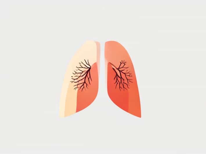
The pleurae are thin, double-layered membranes that surround each lung and line the inside of the thoracic cavity. They play a protective and mechanical role, supporting breathing movement and preventing friction. Even though they are thin, they are incredibly important because they help maintain proper lung expansion and reduce resistance as the lungs inflate and deflate.
Each lung is wrapped in its own pleural sac, meaning there is a separate pleural covering for the right and left lungs. This separation is extremely important because it prevents problems in one lung from automatically affecting the other.
Layers of the Pleurae
The pleurae are divided into two main layers, each with its own position and function but working together as a single system.
The Visceral Pleura
The visceral pleura is the inner layer of the pleurae. It is firmly attached to the surface of the lungs, following every curve, groove, and shape of the lung tissue. Because it is directly in contact with lung tissue, it moves whenever the lungs expand and contract. This close attachment ensures smooth lung motion with minimal friction.
The Parietal Pleura
The parietal pleura forms the outer layer. Unlike the visceral pleura, it does not attach to the lung surface but instead lines the inside of the chest wall, diaphragm, and mediastinum. It is more sensitive to pain and irritation than the visceral layer, which is why pleural inflammation can cause sharp chest pain that worsens with breathing.
The Pleural Cavity Explained
Between the visceral and parietal pleura is a narrow, potential space known as the pleural cavity. Many people imagine it as an empty gap, but in reality, it is filled with a very small amount of pleural fluid. This space and the fluid inside it play a vital mechanical role in breathing.
Even though the cavity is very thin, it is extremely important because it allows the lungs to glide smoothly against the chest wall. Without it, every breath would cause friction, irritation, and damage to delicate lung tissue.
Pleural Fluid and Its Importance
The pleural cavity contains a thin layer of lubricating pleural fluid, which acts like a natural lubricant. This fluid reduces friction between the pleural layers as they slide over each other during breathing movements.
- It prevents friction between the lungs and chest wall
- It allows smooth expansion and contraction of the lungs
- It helps maintain pressure, supporting lung inflation
- It contributes to stability of the lungs within the thoracic cavity
This fluid is constantly produced and reabsorbed in balance. When this balance is disturbed, respiratory problems may develop.
Pressure and Mechanics Inside the Pleural Cavity
The pleural cavity is not only about lubrication. It also helps maintain negative pressure relative to the atmosphere. This negative pressure keeps the lungs expanded and prevents them from collapsing. When you inhale, the chest wall expands, the pleural cavity follows, and the lungs inflate as air fills the airways.
This relationship between pressure, pleural membranes, and lung movement is a key part of respiratory physiology. Without the correct pressure inside the pleural cavity, breathing efficiency would be severely affected.
Different Regions of the Parietal Pleura
The parietal pleura is further divided based on the areas it covers. Understanding these regions helps explain medical descriptions and anatomical explanations.
- Costal pleura Lines the inner surface of the rib cage
- Diaphragmatic pleura Covers the diaphragm
- Mediastinal pleura Covers the mediastinum
- Cervical pleura Extends slightly above the first rib into the root of the neck
Each region adapts to its surrounding structure, maintaining smooth coordination between breathing muscles and the lungs.
Protective Role of the Pleurae
The pleurae are not only mechanical structures; they also provide protection. Because each lung has its own pleural sac, diseases or injury affecting one lung do not automatically spread to the other lung. This separation is crucial for survival in traumatic situations or localized infections.
The pleurae also help filter and drain small amounts of excess fluid through the lymphatic system, maintaining a healthy environment inside the pleural cavity.
Common Conditions Affecting the Pleurae and Pleural Cavity
Although the pleurae are built to function efficiently, they can still be affected by disease, trauma, or infection. Understanding these conditions helps appreciate how delicate and important these membranes are.
Pleurisy or Pleuritis
Pleurisy occurs when the pleura become inflamed. This causes sharp chest pain, especially when breathing deeply, coughing, or moving. The pain happens because the normally smooth pleural surfaces become rough, leading to friction during breathing.
Pleural Effusion
Pleural effusion refers to excess fluid accumulation in the pleural cavity. Too much fluid reduces lung expansion, making breathing difficult. It can occur due to infection, heart problems, injury, or other medical conditions.
Pneumothorax
Pneumothorax happens when air enters the pleural cavity. This disrupts the normal negative pressure, causing the lung to collapse partially or completely. It may result from injury, medical procedures, or spontaneous rupture in some individuals.
Hemothorax
Hemothorax refers to blood collecting inside the pleural cavity, usually due to trauma. Like pleural effusion, it interferes with normal lung expansion.
Why Understanding the Pleurae Matters
Knowing how the pleurae and pleural cavity work helps people better appreciate the complexity of breathing. These thin membranes do more than simply line the lungs. They stabilize, lubricate, protect, and support normal respiratory mechanics. Without them, every breath would be painful and difficult.
For students, healthcare workers, and anyone interested in the human body, understanding this structure strengthens knowledge of respiratory anatomy and helps explain why certain respiratory conditions occur. It also highlights the importance of maintaining lung health and recognizing symptoms related to pleural problems early.
The pleurae and pleural cavity play a silent but powerful role in respiratory function. With two important pleural layers, a carefully maintained pleural cavity, and essential pleural fluid, the lungs are able to move smoothly and efficiently inside the chest. From reducing friction to maintaining pressure and protecting each lung individually, these structures are vital for comfortable breathing. A clear understanding of the pleurae and pleural cavity provides deeper appreciation of how the respiratory system functions every second of life, supporting oxygen supply and overall health.